

Description
Product Overview
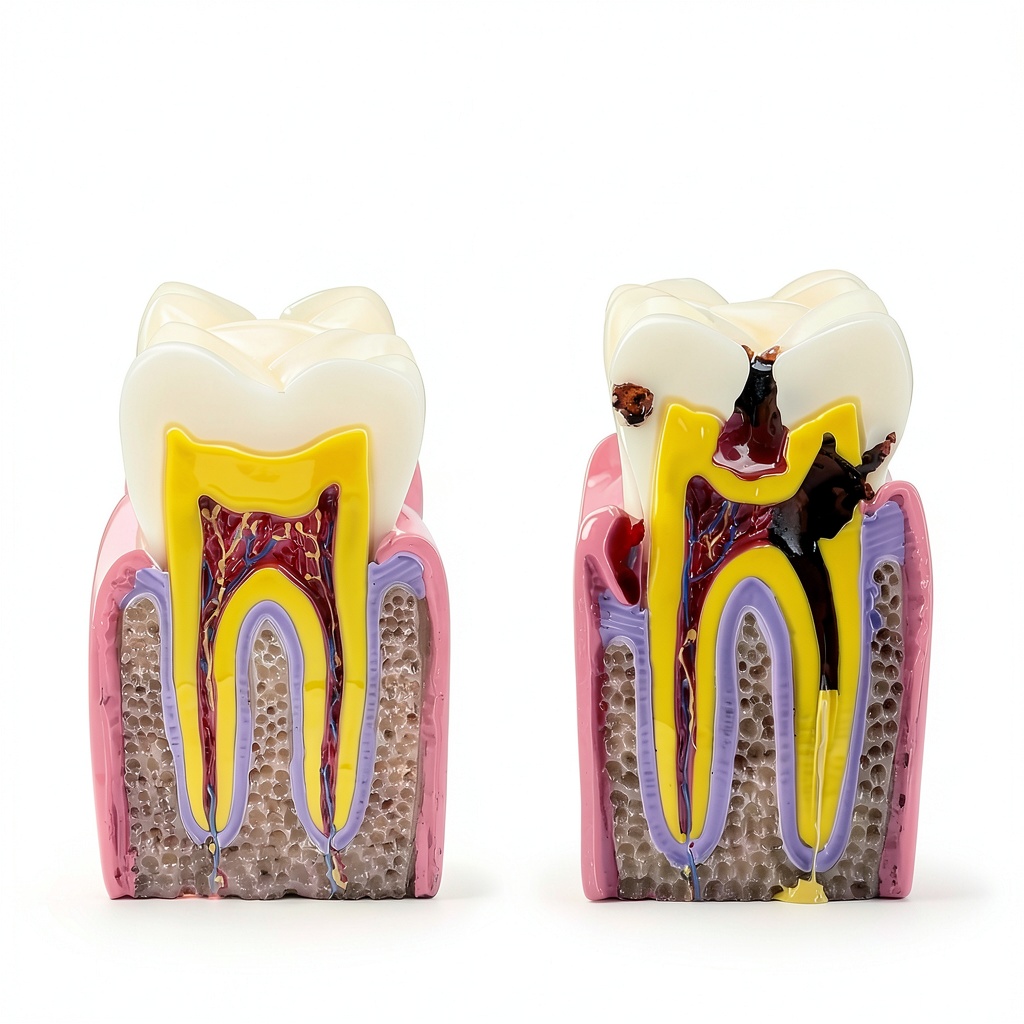
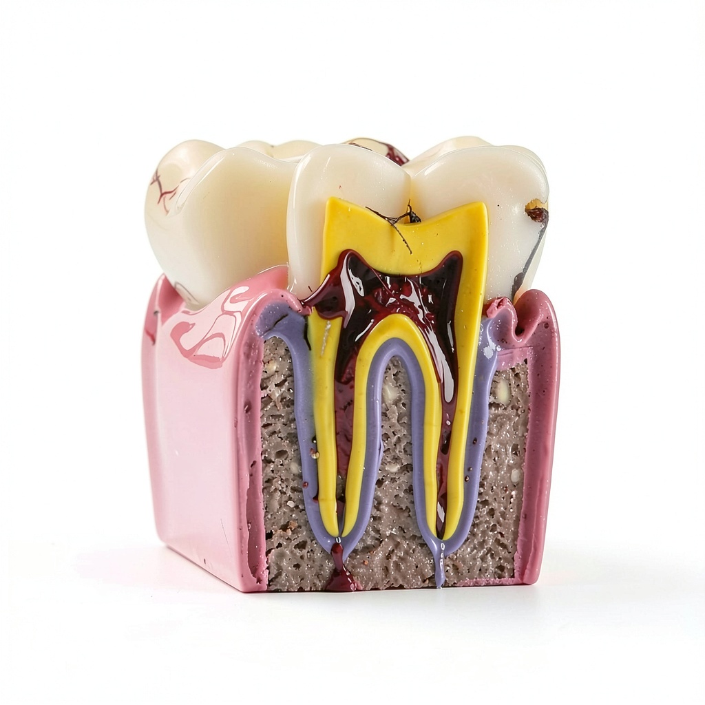
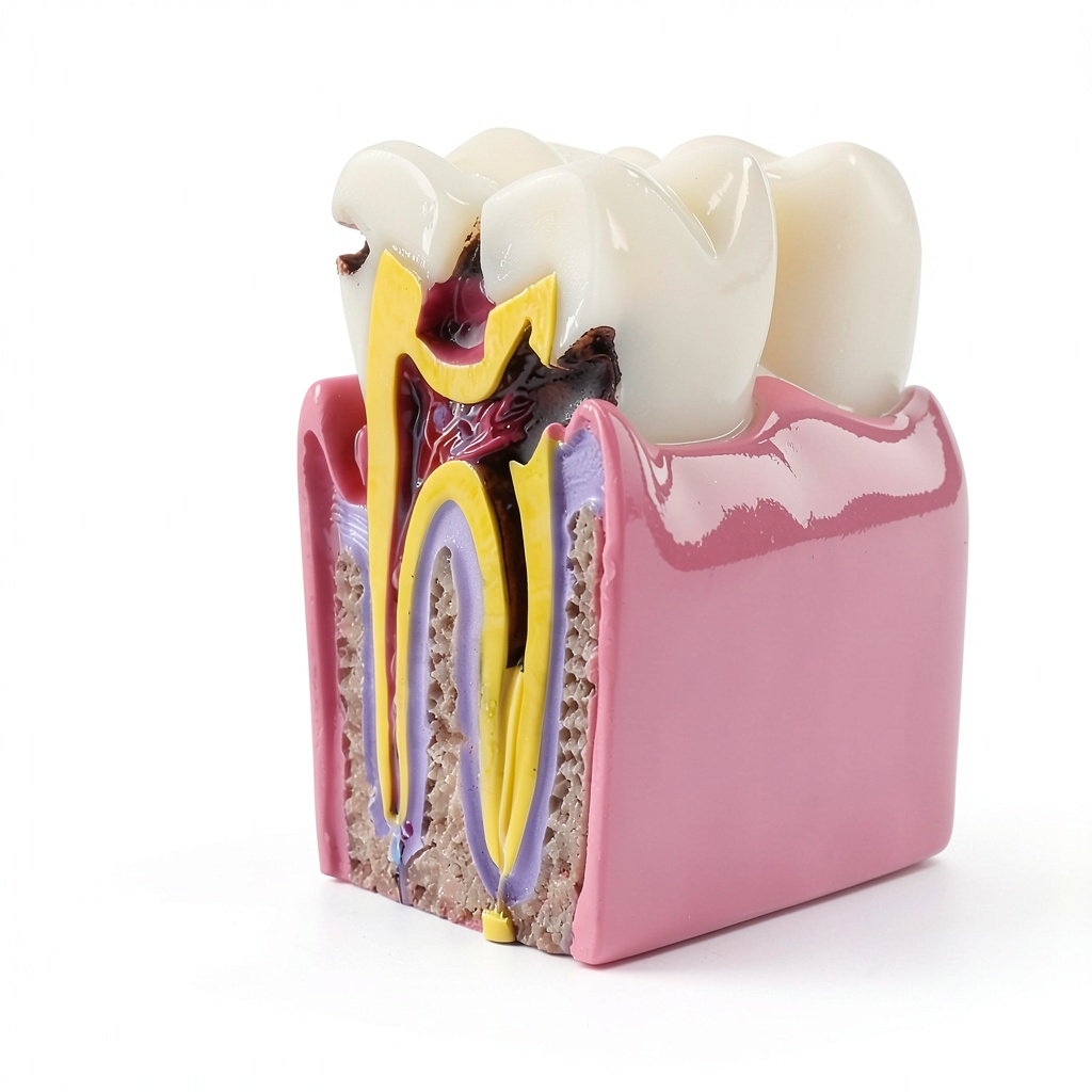
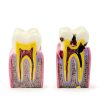
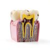
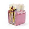
The Alpha Dental 6-times Caries Bilateral Comparison Model is a high‑fidelity educational tool designed to illustrate the development and severity of dental caries on both sides of a tooth. By presenting a side‑by‑side view of healthy and decayed structures, the model enables dentists and educators to demonstrate the visual differences that occur as decay progresses, making complex pathology accessible to patients and students alike. The model’s design incorporates six distinct stages of caries, each accurately scaled to reflect real clinical presentations, allowing a step‑by‑step walkthrough of lesion formation from initial enamel demineralization to deep dentin involvement.
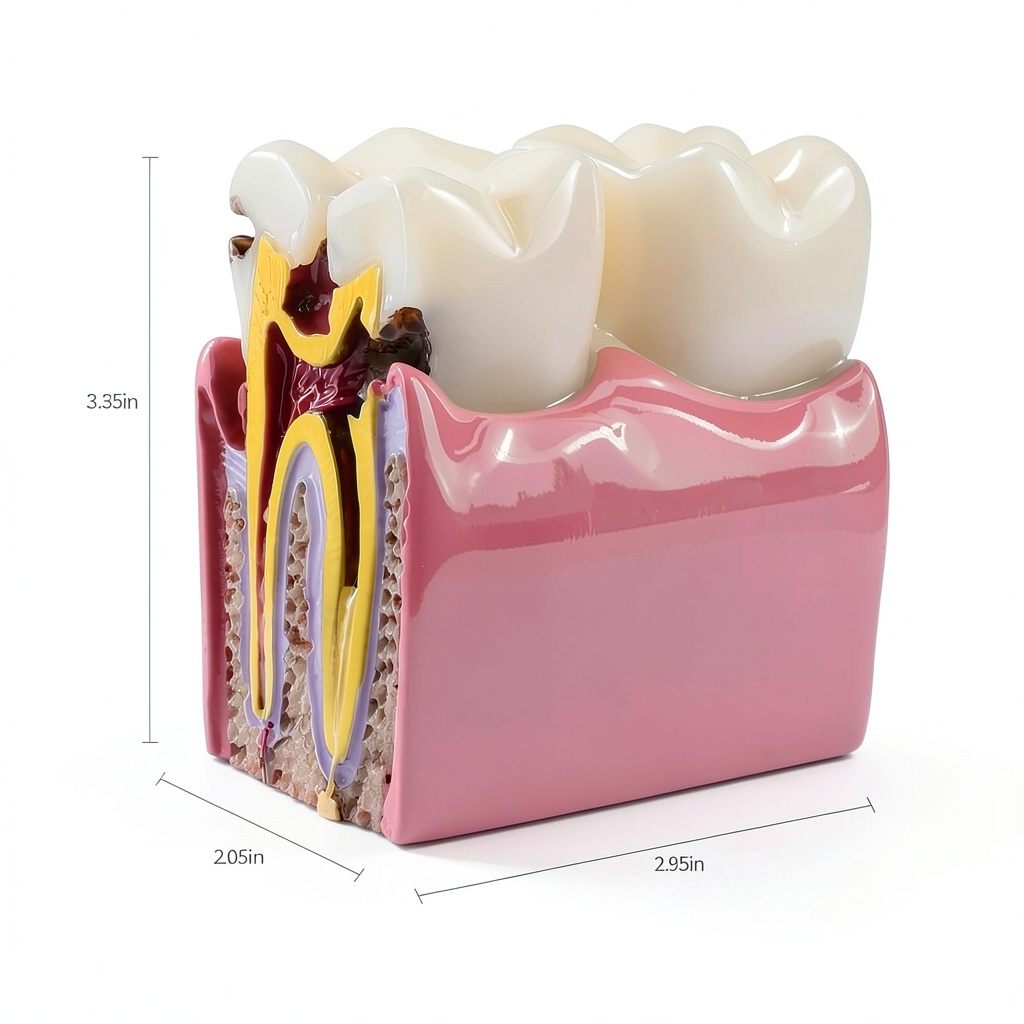
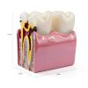
Manufactured from medical‑grade smooth resin, the model offers a lifelike appearance with distinct color differentiation between enamel, dentin, and pulp chambers. The resin is non‑toxic, environmentally friendly, and resistant to wear, ensuring the model remains clear and vibrant even after frequent handling in busy teaching labs. Precise dimensions of 4.13 x 3.54 x 3.11 inches and a lightweight 13.76‑ounce construction make it easy to position on any tabletop or demonstration tray without the need for additional supports. Detailed cross‑sections reveal the exact contours of gum lines, root canals, and internal tooth anatomy, providing a comprehensive view that complements traditional radiographic images.
Beyond its visual accuracy, the model serves as a bridge between theoretical knowledge and practical understanding. When paired with X‑ray images, it offers a three‑dimensional perspective that helps patients grasp the extent of decay, fostering informed consent and encouraging preventive care. Dental schools and continuing education programs benefit from its durability and realistic feel, allowing repeated use in simulations, case studies, and hands‑on workshops. The model’s versatility also extends to research settings, where it can be employed as a standardized reference for comparative studies on caries detection methods.
![[Product front view showing all components]](https://www.distilledco.store/wp-content/uploads/2026/06/0e9d75f872fa46b4a6fb5802b8364cfd.jpg)
Usage
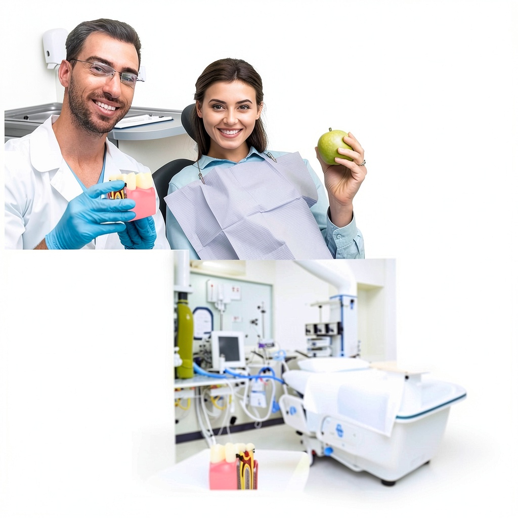
In clinical practice, the model is most effective during patient consultations. Dentists can place the model beside a patient’s intra‑oral photograph or radiograph, pointing out specific areas of concern and explaining how decay progresses through each stage. This visual aid reduces anxiety by demystifying the disease process and clarifies treatment options, whether preventive fluoride applications, minimally invasive restorations, or more extensive restorative procedures.
Educational institutions use the model in lecture halls, simulation labs, and small‑group tutorials. Instructors can demonstrate the impact of oral hygiene habits, dietary choices, and fluoride exposure by referencing the staged caries progression. The model also supports interdisciplinary teaching, allowing dental hygienists, prosthodontists, and oral surgeons to discuss collaborative treatment planning. Its compact size makes it ideal for portable workshops, community outreach events, and patient education kiosks in dental offices.
Why Choose Us
Alpha Dental’s commitment to quality is reflected in every aspect of this model. The resin material is sourced from certified medical suppliers, guaranteeing biocompatibility and long‑term stability. Each unit undergoes rigorous inspection for color consistency, dimensional accuracy, and surface smoothness, ensuring that every educator receives a product that meets professional standards. Our manufacturing process incorporates advanced molding techniques that capture fine anatomical details, from the subtle curvature of the enamel surface to the intricate branching of the root canal system.
Customer support is a cornerstone of our service philosophy. Purchasers receive a dedicated after‑sales liaison who can provide guidance on optimal usage, integration with digital teaching platforms, and troubleshooting any concerns that arise. We also offer a 30‑day satisfaction guarantee and a one‑year warranty against material defects, underscoring our confidence in the model’s durability. By choosing Alpha Dental, institutions gain a reliable, scientifically accurate teaching aid that enhances learning outcomes and supports patient communication.
Key Features
- Six progressive caries stages provide a clear visual timeline for education and patient discussion.
- Medical‑grade smooth resin ensures a realistic look, safe handling, and long‑lasting durability.
- Precise cross‑section reveals enamel, dentin, pulp, and root anatomy for comprehensive study.
- Compact size and lightweight design make it easy to display in any teaching or clinical environment.
- Dedicated after‑sales support and a one‑year warranty give confidence in product performance.
FAQ
What age group is this model suitable for?
The model is designed for use with adults and older children who can understand visual explanations of dental disease. It is especially effective in university dental programs, community health workshops, and private practice settings where patients range from teenagers to seniors.
Can the model be used with digital presentation tools?
Yes, the model can be photographed or scanned and incorporated into PowerPoint slides, virtual simulations, or online patient education portals. Its realistic coloration and detailed anatomy translate well to high‑resolution images for digital use.
Is the resin material safe for repeated handling?
Absolutely. The resin complies with medical safety standards, is non‑toxic, and resistant to scratches and stains. It can be cleaned with mild soap and water, making it ideal for frequent use in busy teaching labs.
How does this model complement traditional X‑ray teaching?
While X‑rays show internal structures in two dimensions, the model adds a three‑dimensional perspective that helps students and patients visualize the spatial relationship of decay to surrounding tooth tissue. Using both tools together creates a more complete educational experience.
What is the return policy if the model arrives damaged?
We offer a 30‑day return window for any damaged or defective units. Contact our support team with photos of the issue, and we will arrange a replacement or refund according to your preference.
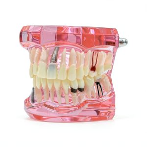
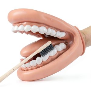 Oral Anatomy Puppet Aiocoid Dental Model Soft Silicone with Toothbrush
  Dinosaur Mystery Kids Book – The Animal Who Series – 35 Page Print
 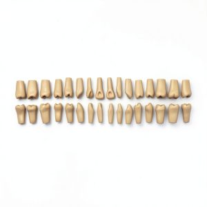 XX Dental Model 32 Ivorine Teeth, Gums, Spring Joint for Columbia 860
  Hydraulic Thumb Kit Wudey for Mini-Excavators 0.8-3 Ton 3-Hole M14-M16
 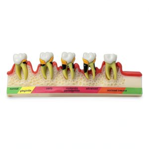 Dentalmall Periodontitis Model 4010 Cross Section Dental Educational
Reviews
There are no reviews yet.